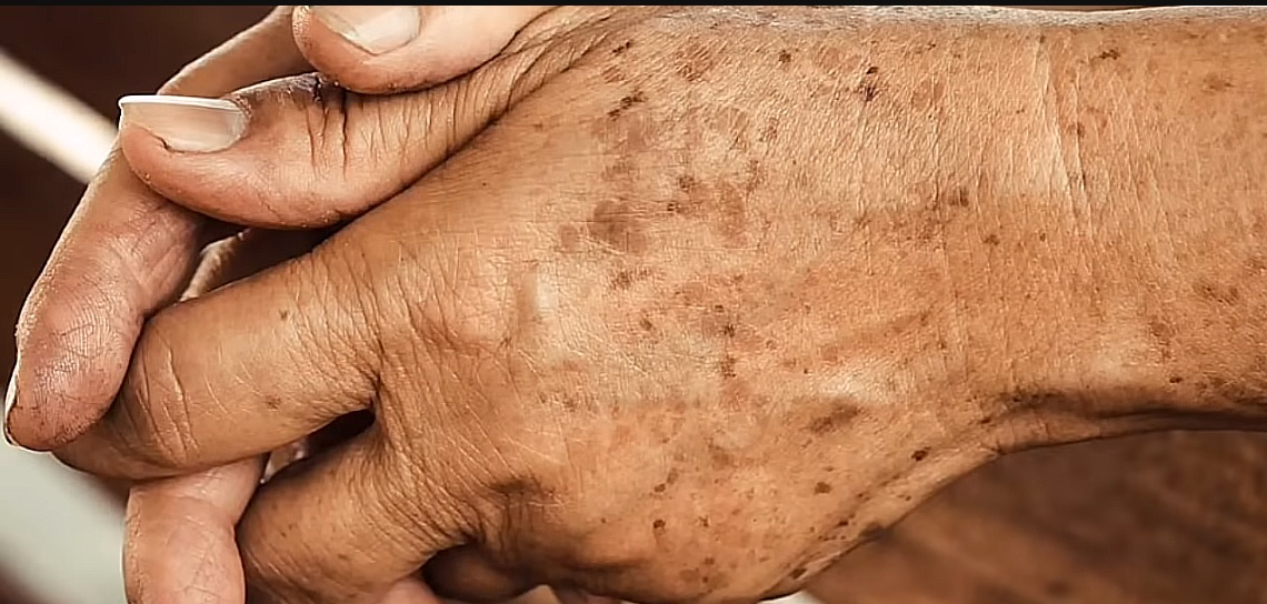
Lipofuscin: The Cell’s Slow-Burn Residue And What It Means For Human Longevity
If aging cells had attic clutter, lipofuscin would be the dusty box you keep meaning to haul out, only to find it has fused to the floor. It is a brownish, autofluorescent gunk that accumulates inside long-lived cells like neurons, cardiac muscle, and the retinal pigment epithelium, resisting normal recycling and quietly rejiggering cell biology over decades. Recent work has moved lipofuscin from harmless bystander to active saboteur, with direct links to oxidative stress, lysosomal failure, and even specific forms of cell death. Here is the state of play, with the most up-to-date evidence and why it matters for longevity science.
What Exactly Is Lipofuscin?
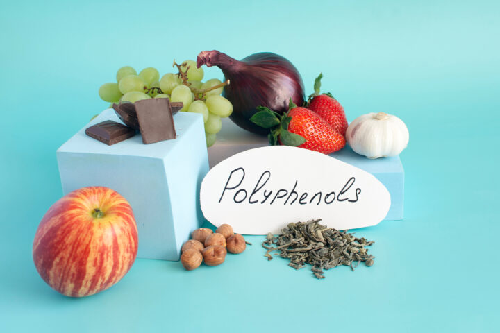
Lipofuscin is a collection of cross-linked proteins and lipids, often laced with reactive by-products of lipid peroxidation and small molecules. In the eye, one major component is the bis-retinoid A2E, which forms from vitamin A chemistry during visual cycling and builds up in the retinal pigment epithelium. Lipofuscin granules are naturally autofluorescent, which is why ophthalmologists can map them with fundus autofluorescence imaging.
Across tissues, lipofuscin tends to accumulate in post-mitotic cells that rarely divide. Human histology shows progressive, intracellular build-up in neurons with age, and ultrastructural studies reveal electron-dense granules packed into cytoplasm. In the retina, age-matched micrographs show RPE cells getting progressively crowded with fluorescent granules from youth to late life.
Marker Or Menace? The New Answer: Both
A key 2024 study used authentic human lipofuscin to test causality. When fibroblasts took it up, lipofuscin triggered mitochondrial reactive oxygen species, destabilized lysosomes, reduced cathepsin D activity, and caused pyroptosis-like cell death at surprisingly low doses. This directly validated a long-suspected lysosome-mitochondria stress loop.
Cardiac biology tells a similar story. Fresh 2025 data show lipofuscin accumulation impairs adult cardiomyocyte function by choking late-stage autophagic flux, a pinpointed mechanism that connects the pigment to contractile decline, not just correlation.
Mechanistically, lipofuscin is not inert. It binds transition metals such as iron and copper, creating a redox-active surface that catalyzes radical formation and further damage, a feed-forward process seen in several tissues with age.
The Retina Is The Proving Ground
The eye offers live access to lipofuscin biology. In the RPE, A2E makes cells unusually sensitive to blue light. New experiments show blue light exposure in A2E-loaded RPE drives ferroptosis, the iron-dependent cell death pathway that thrives on lipid peroxides. That links phototoxic chemistry, lipid peroxidation, and iron to real-world degeneration risk.
Therapeutically, the field has a credible front-runner: remofuscin (soraprazan). It appears to strip pre-existing RPE lipofuscin in animals and primary human cells and has advanced to human studies. A two-year randomized study in Stargardt disease reported slower retinal thinning on SD-OCT with remofuscin tablets versus placebo, and the developer indicates Phase 2 programs for Stargardt and dry AMD. These results are not a panacea, yet they represent the first sustained clinical signal that removing lipofuscin can change retinal structure over time.
Genetic strategies aim upstream. In Stargardt disease, where ABCA4 mutations elevate toxic bis-retinoids, multiple 2024–2025 reviews describe gene or protein-splicing therapies intended to reduce A2E formation at the source, which should secondarily lower lipofuscin load.
Brain, Immune Cells, And Beyond
Lipofuscin builds up in human cortex with age and shows up inside lysosomes of microglia even in young mouse brains, complicating how researchers interpret “engulfment” signals. New mass-spectrometry-based work is mapping the molecular makeup of brain lipofuscin in both normal aging and neuronal ceroid lipofuscinosis, closing the gap between a histological pigment and a defined chemical entity.
The pigment’s reach is systemic. Reviews across 2024–2025 connect lipofuscin to ferroptosis biology in aging tissues and highlight how autofluorescent waste and lysosomal stress are woven through neurodegeneration and other age-related pathologies.
Why Lipofuscin Clogs The Cell’s Cleanup Crew
Lysosomes digest cellular leftovers. Lipofuscin resists their enzymes, and as granules pile up, they dilute lysosomal capacity and blunt autophagy. This is not just theory. Multiple studies show compromised lysosomes limit photoreceptor outer-segment clearance in the RPE and raise lipofuscin-like autofluorescence, while metal-laden lipofuscin fuels oxidant production, turning a recycling center into a slow cooker.
A contemporary 2024–2025 theme is lysosome activation. Boosting TFEB, the master regulator of lysosomal biogenesis, can shrink lipofuscin-like granules in RPE models, and comprehensive lysosome-aging reviews name TFEB and related pathways as logical targets to restore degradative capacity. These are early-stage findings, yet they present a rational route to manage the burden.
Lifestyle Biology Still Matters
Classic work in mammals and newer studies in worms converge on a simple signal: caloric restriction slows lipofuscin accumulation in neurons and other tissues, sometimes alongside cognitive benefits. While CR is not a therapy, it supports the idea that global stress resistance and improved proteostasis can keep the pigment in check.
Light hygiene is sensible for retinal health, since A2E-rich lipofuscin is blue-light sensitive. Laboratory data show blue light worsens oxidative damage and ferroptosis in A2E-loaded RPE cells, reinforcing the eye-care advice that seemed like common sense even before these mechanistic updates.
Metal balance may matter. Given lipofuscin’s love of iron and copper, and growing links between iron handling, lipid peroxidation, and aging, the case for careful iron management continues to strengthen, although clinical guidance must remain conservative until trials connect the dots.
Emerging Toolkits: From Targeted Clearance To Smarter Senolytics
Two complementary strategies are getting attention.
1) Remove or reduce the pigment.
Remofuscin provides human evidence in the eye. Parallel preclinical work suggests mTOR-TFEB activation can reduce lipofuscin-like granules, a logic that could extend to other tissues if delivery hurdles are solved. A 2024 perspective argues that periodic, systemic lipofuscin removal could be a unifying rejuvenation strategy, though this remains a provocative hypothesis rather than clinical fact.
2) Exploit lipofuscin to hit problem cells.
Senescent cells stockpile lipofuscin. A 2025 proof-of-concept built a senolytic that homes to lipofuscin using a binding scaffold linked to a dasatinib payload, increasing selectivity for senescent targets in models. Independent 2025 work also reports that long-term dasatinib plus quercetin can ease lipofuscin-dependent retinal degeneration in mice. Targeting the trash may thus help clear the culprits that make the trash.
Concrete Research Examples You Can Point To
- Authentic lipofuscin triggers damage: Human lipofuscin added to cells caused ROS, lysosomal membrane permeabilization, cathepsin D impairment, and pyroptosis-like death, establishing a direct toxic mechanism.
- Heart cells falter under pigment load: Adult cardiomyocytes with more lipofuscin showed reduced function due to blocked late autophagic steps.
- A2E plus blue light induces ferroptosis: RPE models demonstrate iron-driven lipid peroxidation and death downstream of blue light in A2E-rich cells.
- Clinical signal in humans: The STARTT study reported less retinal thinning over two years in Stargardt patients on oral remofuscin versus placebo. The program is progressing toward Phase 2 for Stargardt and dry AMD.
- Turn up the lysosomes: mTOR-TFEB pathway activation reduced lipofuscin-like autofluorescent granules in RPE models, pointing to a generalizable approach for pigment clearance.
- Dietary restriction slows pigment: From hippocampal neurons in mice to invertebrates, CR regimens lowered lipofuscin burden and correlated with better function.
How To Think About Lipofuscin In Longevity Strategies
1) Treat it as a driver in specific tissues. Evidence in retina and heart now supports causal roles for lipofuscin in dysfunction. That elevates it from passive clock to active lever.
2) Pair upstream prevention with downstream cleanup. Limiting formation of toxic precursors, improving proteostasis and lysosomal capacity, and, where possible, removing existing deposits is a coherent plan. RPE work leads here, with applicability to brain and heart as delivery technologies improve.
3) Watch iron and light in the eye. Emerging ferroptosis links and A2E phototoxicity suggest practical modifiers, from blue-light management to thoughtful iron biology, while the field refines targeted therapeutics.
What To Watch Next
- Phase 2 remofuscin readouts and whether structural benefits translate to preserved vision and measurable lipofuscin reduction in humans.
- TFEB-directed therapies that safely scale lysosomal biogenesis in human tissues beyond the eye.
- Ferroptosis modulators as protectants in A2E-rich, iron-loaded environments like the RPE.
- Senolytics that piggyback on lipofuscin to find their targets with higher precision.
- Deeper composition maps of brain and heart lipofuscin to guide enzyme or binding-based removal strategies.
References
- Baldensperger, T., Dominici, S., Meehan, B., Toprakcioglu, Z., Kogelmann, J., Milenkovic, V. M., Solanki, R., Krahmer, N., Kosyakova, N., Plockinger, U., Holmgren, A., Ziegler, D. V., Desel, C., von Haeseler, A., & Arlt, A. (2024). The age pigment lipofuscin causes oxidative stress, lysosomal dysfunction, and pyroptotic cell death. Free Radical Biology and Medicine, 225, 871–880. https://doi.org/10.1016/j.freeradbiomed.2024.10.311
- Walter, S., Leonhartsberger, S., Wieser, K., Käß, M., Weninger, A., Feichtinger, R., Hofer, D., Daga, M., Sabirsh, A., & Cougnoux, A. (2025). Oxidized protein aggregate lipofuscin impairs cardiomyocyte function by diminishing late-stage autophagic flux. Redox Biology, 81, 103559. https://doi.org/10.1016/j.redox.2025.103559
- Yang, B., Wu, Y., Zhang, J., Pan, H., Xu, J., Han, H., & Li, J. (2025). Exposure of A2E to blue light promotes ferroptosis in the retinal pigment epithelium. Cellular & Molecular Biology Letters, 30, 22. https://doi.org/10.1186/s11658-025-00700-2
- Dhooge, P. P. A., Möller, P. T., Boon, C. J. F., van Genderen, M., Meester-Smoor, M., Long, V., Mohand-Said, S., Sahel, J.-A., Leroy, B. P., & Hoyng, C. B. (2022). The STArgardt Remofuscin Treatment Trial (STARTT): Design and baseline characteristics of enrolled Stargardt patients. Open Research Europe, 1, 96. https://doi.org/10.12688/openreseurope.13872.3
- Katairo GmbH. (2023, September 5). Katairo reports positive outcomes from its Phase 2 STARTT study with Remofuscin in Stargardt disease [Press release]. https://katairo.com
- Höhn, A., Jung, T., Grimm, S., & Grune, T. (2010). Lipofuscin-bound iron is a major intracellular source of oxidants: Role in senescent cells. Free Radical Biology and Medicine, 48(8), 1100–1108. https://doi.org/10.1016/j.freeradbiomed.2010.01.030
- Moore, W. A., Davey, V. A., Weindruch, R., Walford, R., & Ivy, G. O. (1995). The effect of caloric restriction on lipofuscin accumulation in mouse brain with age. Gerontology, 41(Suppl. 2), 173–185. https://doi.org/10.1159/000213741
- Guan, Z., Cai, B., Zhang, H., Lin, C., Abraham, A., & Sparrow, J. R. (2020). A2E distribution in RPE granules in human eyes. Molecules, 25(6), 1413. https://doi.org/10.3390/molecules25061413
- Stillman, J. M., Hsu, E. T., & Stevens, B. (2023). Lipofuscin-like autofluorescence within microglia and its impact on studying microglial engulfment. Nature Communications, 14, Article 6969. https://doi.org/10.1038/s41467-023-42809-y
- Julien-Schraermeyer, S., Illing, B., Tschulakow, A. V., & Schraermeyer, U. (2020). Penetration, distribution, and elimination of remofuscin (soraprazan) in rats and monkeys. Pharmacology Research & Perspectives, 8(4), e00683. https://doi.org/10.1002/prp2.683
- Bourauel, L., Schweitzer, D., Hammer, M., & Paetzold, J. (2024). Spectral analysis of human retinal pigment epithelium cells using fluorescence lifetime imaging ophthalmoscopy. Investigative Ophthalmology & Visual Science, 65(1), 10. https://doi.org/10.1167/iovs.65.1.10
- Zhao, N., Jiang, Y., Zhou, Y., & Li, X. (2025). Ferroptosis: An energetic villain of age-related macular degeneration. Aging and Disease, 16(2), 365–382. https://doi.org/10.14336/AD.2025.0141
- Lyu, J., Wang, Z., Hou, Y., Chen, L., & Xu, Y. (2021). Iron accumulation and lipid peroxidation in the aging retina: Implication of ferroptosis in age-related macular degeneration. Aging and Disease, 12(2), 529–551. https://doi.org/10.14336/AD.2020.0912